MicroscopeLenses are no longer necessary for some microscopes, according to the engineers developing FlatScope, a thin fluorescent microscope whose abilities promise to surpass those of old-school devices.

A paper in Science Advances describes a wide-field microscope thinner than a credit card, small enough to sit on a fingertip, and capable of micrometer resolution over a volume of several cubic millimeters.

FlatScope eliminates the tradeoff that hinders traditional microscopes in which arrays of lenses can either gather less light from a large field of view or gather more light from a smaller field.

Rice University engineers Ashok Veeraraghavan, Jacob Robinson, Richard Baraniuk, and their labs began developing the device as part of a federal initiative by the Defense Advanced Research Projects Agency as an implantable, high-resolution neural interface. But the device’s potential is much greater.

(more…)

Nanoscale Microscopy

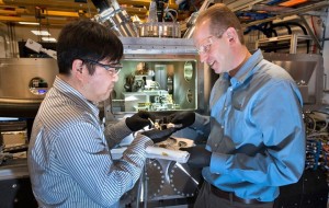
The microscope they developed produces x-ray images by scanning a sample while collecting various x-ray signals emerging from the sample.Image: Brookhaven National Laboratory

The microscope they developed produces x-ray images by scanning a sample while collecting various x-ray signals emerging from the sample.
Image: Brookhaven National Laboratory

Researchers have developed a new x-ray microscope that will provide scientists with the opportunity to image nanostructures and chemical reactions down to the nanometer.

The new class of x-ray microscope allows for nanoscale imagining like never before. This development brings researchers one step closer to the ultimate goal of nanometer resolution.

This from Brookhaven National Laboratory:

The microscope manipulates novel nanofocusing optics called multilayer Laue lenses (MLL) — incredibly precise lenses grown one atomic layer at a time — which produce a tiny x-ray beam that is currently about 10 nanometers in size. Focusing an x-ray beam to that level means being able to see the structures on that length scale, whether they are proteins in a biological sample, or the inner workings of a fuel cell catalyst.

(more…)

Amazing Microscopy Videos and Images

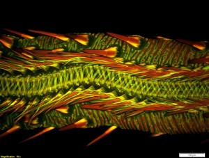
The Olympus BioScapes competition is held to celebrate the intersection of art and science.Credit: Dr. Matthew S. Lehnert of Kent State University at Stark

The Olympus BioScapes competition is held to celebrate the intersection of art and science.
Credit: Dr. Matthew S. Lehnert of Kent State University at Stark

We sometimes get so wrapped up in the technicality of science that we forget how beautiful it can be. Microscopy in particular provides us with the ability to see remarkable worlds that are otherwise invisible to the naked eye.

The Olympus BioScapes competition is held every year to help celebrate the intersection of art and science. Scientists from around the world submit their photos and videos of microscopy work to be judged “based on the science they depict, their beauty or impact, and the technical expertise involved in capturing them.”

(more…)

The Art of Dried Whiskey and Microscopy

The image to your right may look like a fine art print of an ocean scene at night, but it’s actually just a close-up of some dried Glenlivet 162, or for those of you who aren’t avid alcohol connoisseurs – it’s simply a photo of whiskey.

Maybe “simple” is not the best word to describe the chemical process that takes place, but the discovery that whiskey can make these beautiful images had a humble beginning.

Professional artist and photographer Ernie Button started creating photos of the patterns formed after letting a drop or two of whiskey dry at the bottom of a glass, which resulted in these clear and rhythmic images.

Though he loved the aesthetic value, Button wanted to understand why the images looked the way they looked.

(more…)

Van Gogh under the Microscope

By examining paint segments from Van Gough's "Sunflowers," experts believe preservation techniques could be improved.Credit: Van Gogh Gallery

By examining paint segments from Van Gogh’s “Sunflowers,” experts believe preservation techniques could be improved.
Credit: Van Gogh Gallery

Electrochemical and solid state science transcend the limits of academic science to touch many of the things we come into contact with on a day-to-day basis, whether we know it or not. Most recently we’ve gotten a first-hand account of this at our Electrochemical Energy and Water Summit, where some of the brightest minds in electrochemical and solid state science came together to solve critical issues in global sanitation. Now, these sciences are even assisting in the preservation of culture.

Pin-sized painting samples from Vincent van Gogh’s “Sunflowers” painting have been extracted from the Van Gogh Museum and are now under the microscope at The University of Queensland’s Centre for Microscopy and Microanalysis (CMM).

UQ’s Professor John Drennan is leading the project, which aims to understand the aging characteristics of significant artworks in order to improve conservation techniques.

(more…)

New Microscope Collects Dynamic Images

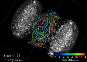
Growing microtubule endpoints and tracks are color coded by growth phase lifetime.Credit: Betzig Lab, HHMI/Janelia Research Campus, Mimori-Kiyosue Lab, RIKEN Center for Developmental Biology

Growing microtubule endpoints and tracks are color coded by growth phase lifetime.
Credit: Betzig Lab, HHMI/Janelia Research Campus, Mimori-Kiyosue Lab, RIKEN Center for Developmental Biology

A new discovery out of Howard Hughes Medical Institute’s Janelia Research Campus is allowing biologists to see 3-D images of subcellular activity in real time.

They’re calling it lattice light sheet microscopy, and it’s providing yet another leap forward for light microscopy. The imaging platform was developed by Eric Betzig and colleagues in order to collect high-resolution images rapidly and minimize damage to cells.

Continue reading to check out the amazing video that shows the five different stages during the division of a HeLa cell as visualized by the lattice light sheet microscope.

(more…)

The 2014 Nobel Prize in Chemistry has been awarded “for the development of super-resolved fluorescence microscopy.”

The awardees are as follows: Eric Betzig, 54, of the Howard Hughes Medical Institute’s Janelia Research Campus in Ashburn, VA.; Stefan W. Hell, 51, of the Max Planck Institute for Biophysical Chemistry in Gottingen, and the German Cancer Research Center in Heidlberg, Germany; and William E. (W.E.) Moerner, 61, of Stanford University.

Because of these men, it is possible for us to obtain optical images at the nanometer scale and understand molecules.

This from The Royal Swedish Academy of Sciences:

For a long time optical microscopy was held back by a presumed limitation: that it would never obtain a better resolution than half the wavelength of light. Helped by fluorescent molecules the Nobel Laureates in Chemistry 2014 ingeniously circumvented this limitation. Their ground-breaking work has brought optical microscopy into the nanodimension.

Read the full article here.

The committee noted the importance of these achievements, by which we’re able to see how proteins in fertilized eggs divide into embryos and track proteins involved in Alzheimer’s or Parkinson’s disease.

“Due to their achievements, the optical microscope can now peer into the nanoworld,” said the committee.

Find out more information on the discoveries of the winners at Chemical & Engineering News.

Also, check out our past issue of Interface entitled, “25 Years of Scanning Electrochemical Microscopy.” The journal is completely open access, allowing everyone to partake in and share this wealth of information.