
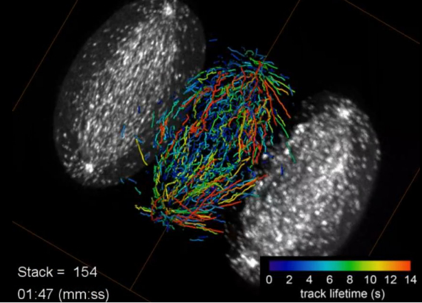
Growing microtubule endpoints and tracks are color coded by growth phase lifetime.
Credit: Betzig Lab, HHMI/Janelia Research Campus, Mimori-Kiyosue Lab, RIKEN Center for Developmental Biology
A new discovery out of Howard Hughes Medical Institute’s Janelia Research Campus is allowing biologists to see 3-D images of subcellular activity in real time.
They’re calling it lattice light sheet microscopy, and it’s providing yet another leap forward for light microscopy. The imaging platform was developed by Eric Betzig and colleagues in order to collect high-resolution images rapidly and minimize damage to cells.
Continue reading to check out the amazing video that shows the five different stages during the division of a HeLa cell as visualized by the lattice light sheet microscope.