Image: NYU’s Jerschow Lab
Researchers from New York University have developed a new technique to give a highly detailed, 3D look inside a lithium-ion battery.
“One particular challenge we wanted to solve was to make the measurements 3D and sufficiently fast, so that they could be done during the battery charging cycle,” explains Alexej Jerschow, co-author of the study that details the development. “This was made possible by using intrinsic amplification processes, which allow one to measure small features within the cell to diagnose common battery failure mechanisms. We believe these methods could become important techniques for the development of better batteries.”
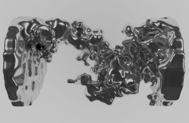
The look that the researchers offer gives new insight to dendrites – the deposits that build up inside a Li-ion battery that can affect performance and safety. To do this, the team used MRI technology to focus the image and took an additional step to improve image quality.
This from New York University:
The researchers sought to enhance this process by focusing on the lithium’s surrounding electrolytes—substances used to move charges between the electrodes. Specifically, they found that MRI images of the electrolyte became strongly distorted in the vicinity of dendrites, providing a highly sensitive measure of when and where they grow.
“The method examines the space and materials around dendrites, rather than the dendrites themselves,” says Andrew Ilott, lead author of the paper. “As a result, the method is more universal. Moreover, we can examine structures formed by other metals, such as, for example, sodium or magnesium—materials that are currently considered as alternatives to lithium. The 3D images give us particular insights into the morphology and extent of the dendrites that can grow under different battery operating conditions.”

