The Olympus BioScapes competition is held to celebrate the intersection of art and science.
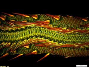
Credit: Dr. Matthew S. Lehnert of Kent State University at Stark
We sometimes get so wrapped up in the technicality of science that we forget how beautiful it can be. Microscopy in particular provides us with the ability to see remarkable worlds that are otherwise invisible to the naked eye.
The Olympus BioScapes competition is held every year to help celebrate the intersection of art and science. Scientists from around the world submit their photos and videos of microscopy work to be judged “based on the science they depict, their beauty or impact, and the technical expertise involved in capturing them.”
This year’s Olympus BioScape winners have just been announced.
A group of researchers who used a novel imaging technique to track a cell in a developing fly embryo took first prize. The video may help reveal cell lineages, cell differentiation and whole-embryo morphogenesis. Take a look at the video below:
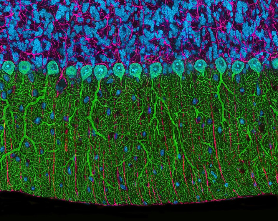
Second prize went to a researcher from the National Center for Microscopy and Imaging Research at the University of California for his microscopic image of a rat brain cerebellum using multiphoton photography.
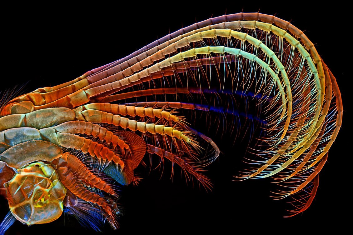
Coming in third is Dr. Igor Siwanowicz of HHMI Janelia Research Campus in Ashburn, VA for his image of barnacle appendages that sweep plankton and other food into the barnacle’s shell for consumption.
The researchers at HHMI Janelia Research Campus also grabbed another top ten spot with their 3-D recordings of the brain signals from 100,000 neurons. Video below:
The Max Planck Institute also made the list with this 3-D reconstruction of a zebrafish’s beating heart in this video:
Make sure to check out the gallery to see the rest of the winners and honorable mentions!
And if you’re into microscopy, make sure to check out “25 Years of Scanning Electrochemical Microscopy” from the Summer 2014 issue of Interface – it’s Open Access!


